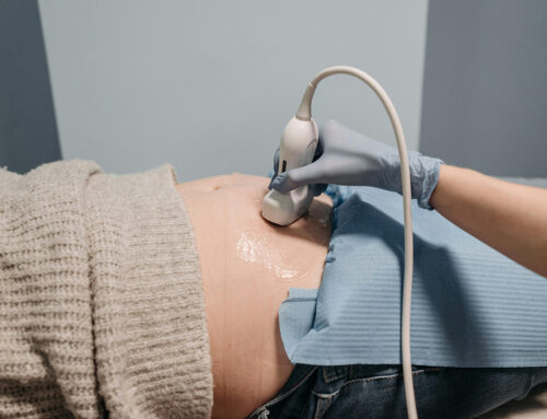
The Significance of Ultrasound as Part of a Natural Birthing Plan
Pregnancy is a journey of transformation and discovery, where each step brings a deeper connection between mothers and their unborn children. In the United States, ultrasounds have become a significant part of this journey, providing expectant mothers and their families with an invaluable tool for health monitoring and emotional bonding.
When you choose midwifery care, you might wonder about ultrasounds during pregnancy. Use of this technology has become increasingly common in obstetrics, with the vast majority of pregnant people choosing to have ultrasounds. With ultrasounds becoming increasingly overused in maternity care, midwives encourage shared decision making when it comes to ultrasounds, and tailor ultrasound frequency to the need and desire of the family. Generally speaking, obstetric ultrasounds are used in midwifery care to help identify pregnancy complications and possible high-risk pregnancies that may not be able to be confirmed with a physical clinical examination.

The Current Trends
Ultrasound technology has been embraced widely across the U.S., offering a non-invasive peek into the womb. The American College of Obstetricians and Gynecologists (ACOG) recommends that all expectant parents have at least one ultrasound during pregnancy for fetal anatomy, usually between 18 and 22 weeks. While many people consider frequent ultrasounds to be standard during pregnancy, the American College of Nurse-Midwives (ACNM) and the National Association of Certified Professional Midwives caution providers about the overuse of the technology.
The Technology
An ultrasound works by sending high-frequency sound waves through the uterus. These sound waves bounce off the baby, creating echoes that are converted into visual images of the baby’s shape and position in the womb. Diagnostic ultrasound is generally regarded as safe and do not produce ionizing radiation like that produced by x-rays.
The Benefits
The benefits of ultrasounds extend beyond the physical health monitoring of the baby. They provide a first “picture” of the baby, often strengthening the emotional bond between the child and parents. For many families, seeing the baby on the ultrasound screen is the moment pregnancy feels “real.” It’s also a reassuring experience, as parents can visually confirm the healthy development of their child. Furthermore, ultrasounds can help families plan for any medical needs the baby might have after birth.
How Ultrasounds Support Natural Births
For those considering natural, out-of-hospital births, such as those facilitated at birthing centers like South Coast Midwifery, ultrasounds can be particularly supportive. At birthing centers, the focus is on minimizing medical intervention while ensuring the safety and health of both mother and baby. Ultrasounds can help by providing detailed information about the baby’s fetal anatomy, and position, which is crucial for anticipating the needs during a natural birth.
How Many Ultrasounds Do You Need
The number of ultrasounds can vary based on medical need, personal preference, and healthcare provider recommendations. Typically, a low-risk pregnancy might include anywhere from one to three to four scans. If a first trimester ultrasound is indicated, it would ideally be performed around 8 to 12 weeks to confirm the pregnancy and estimate or corroborate a due date. Further scans at around 18 to 20 weeks are used to assess fetal anatomy and development. Additional scans may be recommended to monitor specific aspects of health and development as the pregnancy progresses, such as position, growth discrepancies, placenta complications, etc.
The 20-Week Ultrasound
The 20-week ultrasound, often known as the anatomy scan, holds particular importance. It’s not only about confirming the baby’s gender, if desired; this scan provides a detailed check of the baby’s organs and systems. It assesses the growth and development of the baby and can help in detecting any anomalies early. It also examines the placenta’s health and position, crucial for anticipating complications during birth. This ultrasound serves as a critical checkpoint for many parents, providing peace of mind and the joy of seeing a more defined image of their baby. Most women in the U.S. will experience several ultrasound scans during their pregnancy, but if a family wishes to limit them, sometimes the anatomy scan may be the only one recommended or needed. This session ensures the health and development of the baby, and gives reassurance that an out-of-hospital birth is a safe option.
Ultrasound technology has woven itself into the fabric of prenatal care, offering a blend of health monitoring and emotional support. Over the years, advancements in ultrasound technology have made these images clearer, thus enriching the experience for expectant parents. As technology advances and becomes even more integrated into family-centered care, its role in enriching the pregnancy experience continues to grow, but as with any form of technology, the risk of diagnostic errors can exist. Overdiagnosis, underdiagnosis, and inaccurately reported test findings are all examples of diagnostic mistakes. False-positive results may prompt unneeded diagnostic testing, undue stress and even therapeutic interventions. For that reason, in low-risk pregnancies, it is also completely safe to limit ultrasounds and use them only when clinically indicated to support a midwife’s physical examinations and assessments.


